Summary for recent publications
2015
The destiny of Ca2+ released by mitochondria.
Takeuchi A, Kim B, Matsuoka S.
J Physiol Sci. 65(1), 11-24, 2015. doi:10.1007/s12576-014-0326-7
Mitochondria have long been known as ATP producing factories as well as Ca2+
stores. Since heart is one of the organs which generate and utilize a huge
amount of ATP, and in which repetitive Ca2+ transients occur, we
have been studyingon the coupling of mitochondrial and cellular functions in the
heart. Using guinea-pig ventricular myocytes, we found that the increase of
stimulus frequency dramatically increased the mitochondrial Ca2+ as
well as NADH, which suggests that mitochondrial Ca2+changes
dynamically and is involved in the regulation of energy metabolism in the heart
(Jo et al., J Mol Cell Cardiol, 40:394-404, 2006).The dynamics of mitochondrial
Ca2+ is determined by the influx via Ca2+uniporter (MCU)
and the efflux via Na+-Ca2+ exchanger and/or Ca2+-H+
exchanger. It has been well recognized that the Ca2+ efflux out of
mitochondria in the heart is mainly via Na+-Ca2+
exchanger.However, the stoichiometry (ion-exchange ratio) and the
electrogenicityof the mitochondrial Na+-Ca2+ exchanger were
controversial.We clearly demonstrated, by use of permeabilized rat ventricular
myocytes, that the mitochondrial Na+-Ca2+ exchanger is
voltage dependent and electrogenic, which suggests the stoichiometry is >3Na+
for 1Ca2+ (Kim et al.,J Physiol,
586:1683-1697, 2008).Since the identification of NCLX as a mitochondrial Na+-Ca2+
exchanger in 2010 (Paltyet al., ProcNatlAcadSci USA, 107:436-441, 2010),
we have been focusing on the functional characterization of NCLX in B
lymphocytes and cardiomyocytes and reported that NCLX is the major Ca2+
extruder out of mitochondria in these cells (Kim et al., J Physiol,
590:459-474, 2012; Kim et al., AdvExp Med Biol, 961:19-201, 2013). In
2013, we found that NCLX is involved in regulating the automaticity of HL-1
cell line originating from mouse atrial myocytes (Takeuchi et al., Sci Rep, 3:2766,
2013).Knocking down NCLX by use of siRNA dramatically slowed down the
generation of automaticity (Fig. A). In addition, NCLX knockdown decelerated
the sarcoplasmic reticulum Ca2+ reuptake rate (Fig. B), suggesting
that NCLX participates in the Ca2+ crosstalk between mitochondria and sarcoplasmic reticulum.The mechanisms
underlying the NCLX reduction-mediated slowing of automaticity were analysed
using our own-made mathematical model of HL-1 cells. It was indicated that
automaticity of HL-1 cells is determined by spontaneous Ca2+ leak from sarcoplasmic
reticulum. Simulation of NCLX reduction showed that Ca2+ supply from
the mitochondria to the sarcoplasmic reticulum decreased to slow down the
firing rate. The timing of Ca2+ induced Ca2+ release,
activation of the inward current of the plasma membrane Na+-Ca2+
exchanger (NCX), and thus the timing of activation of voltage-dependent Na+
channel (Nav1) and voltage-dependent Ca2+ channel (VDCC) was delayed, resulting in prolongation of the cycle length (Fig. C). Taken together, our combined experiments and simulations indicated that NCLX regulates the rhythmicity of HL-1 cells via crosstalk between mitochondria and sarcoplasmic reticulum. 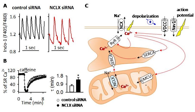
2014
Rat Uterine Oxytocin Receptor and Estrogen Receptor α and β mRNA Levels
are Regulated by Estrogen through Multiple Estrogen Receptors.
Murata T, Narita K and Ichimaru T.
J Reprod Dev. 2014; 60:55
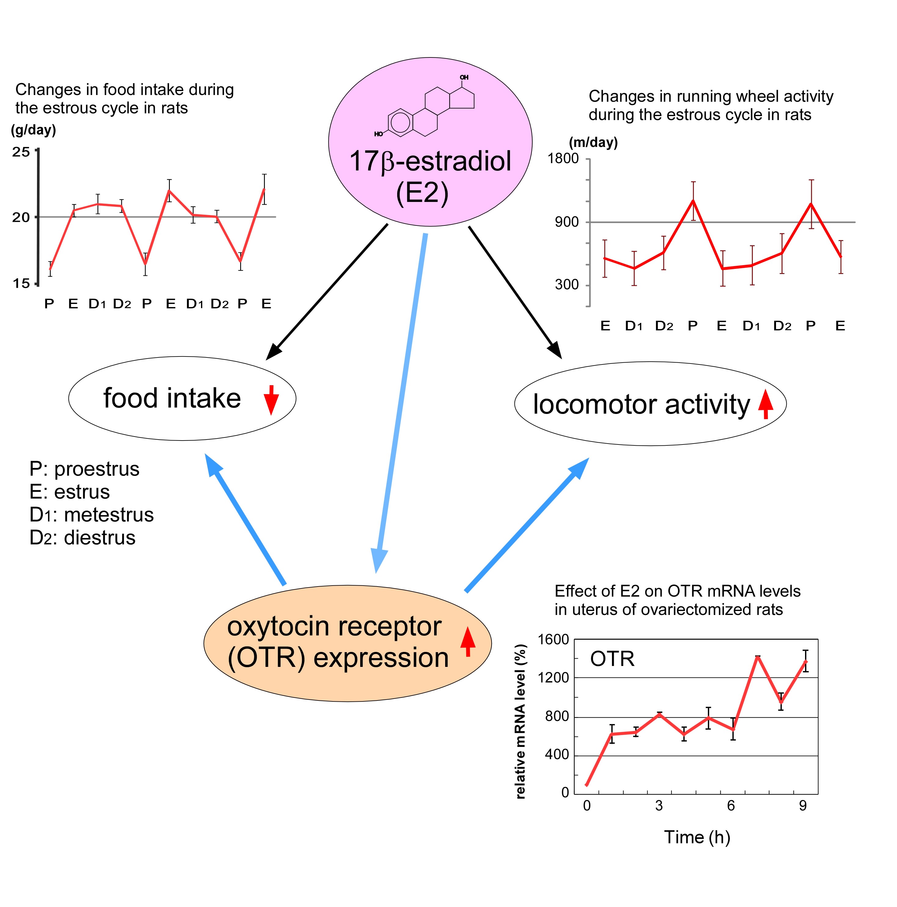
Oxytocin was initially isolated as a neurohypophysial hormone that stimulates
contraction of the myometrium and myoepithelium to facilitate parturition
and milk ejection, respectively. It has also been shown to play a role
in various reproductive functions in the mammary gland, ovary, brain and
uterus. In the uterus, the near-term myometrium is extremely sensitive
to oxytocin. This increased uterine responsiveness to oxytocin was shown
to occur in parallel with an increase in uterine oxytocin receptor (OTR)
mRNA expression in several animals including rats [Liu et al., J Endocrinol
1996; 150:479]. Although estrogen stimulates an increase in OTR mRNA expression
in ovariectomized (OVX) virgin rats, an injection of estrogen did not stimulate
OTR mRNA expression in late pregnant rats or in progesterone-primed OVX
virgin rats and was only effective following ovariectomy and the removal
of progesterone, respectively [Murata et al., J Endocrinol 2000; 166:45].
These results suggest that, in addition to increases in serum estrogen
levels in near-term rats, the regulation of uterine responsiveness to estrogen
is an important part of the mechanism of action of estrogen in the uterus.
Estrogen action is mediated through several types of receptors (ERs), such
as ERα, ERβ and putative membrane ERs. However, which types of ERs are
involved in OTR expression has not been elucidated. Therefore, the effects
of 17β-estradiol, an ERα agonist (PPT), an ERβ agonist (DPN) or a selective
estrogen receptor modulator estren (Es) on uterine OTR, ERα and ERβ levels
were examined in OVX rats [Murata et al., J Reprod Dev 2014; 60:55]. This
study suggests that uterine OTR gene expression is upregulated by estrogen
through the classical nuclear (or non-nuclear) ERs, ERα and ERβ, while
the levels of these ERs are downregulated by estrogen through multiple
pathways including Es-sensitive nonclassical ERs.